Wednesday 29 March 2023

Patellofemoral Pain Syndrome (PFPS) : Cause, Symptoms, Treatment

What is Patellofemoral pain Syndrome?

Patellofemoral pain Syndrome (PFPS)

Patellofemoral pain syndrome (PFPS) is a condition characterized by pain in the front of the knee, around the kneecap (patella), and the surrounding structures. It is a common condition that affects athletes and active individuals, especially those who participate in sports that involve repetitive knee bending, such as running, jumping, and squatting.

PFPS is usually caused by an imbalance in the muscles that support the knee, such as the quadriceps, hamstrings, and hip muscles. This imbalance can cause the patella to track abnormally over the femur, leading to irritation and inflammation of the soft tissues around the patella.

Symptoms of PFPS include pain in the front of the knee, especially during activities that involve bending the knee, such as going up and down stairs, squatting, or kneeling. The pain may be worse after sitting for prolonged periods or during activities that involve prolonged periods of standing or walking.

Treatment of PFPS typically involves rest, ice, and physical therapy to correct muscle imbalances and improve joint mechanics. In some cases, bracing or taping of the knee may be recommended to help support the patella and reduce pain. Severe cases may require surgery, but this is rare.

Related Anatomy

To understand patellofemoral pain syndrome (PFPS), it's helpful to have a basic understanding of the anatomy of the knee joint and surrounding structures.

The knee joint is the largest joint in the body and is made up of three bones: the femur (thigh bone), the tibia (shin bone), and the patella (kneecap). The patella sits in a groove at the end of the femur and helps to transmit the force of the quadriceps muscle to the lower leg bones.

The quadriceps muscle is the large muscle group on the front of the thigh that extends the knee. It is made up of four muscles that come together to form a tendon that attaches to the patella. The patellar tendon attaches the patella to the tibia, and helps to transmit the force of the quadriceps muscle to the lower leg bones.

The knee joint is surrounded by ligaments, which connect the bones together and provide stability. The medial and lateral collateral ligaments are on the sides of the knee, while the anterior and posterior cruciate ligaments are inside the joint and help to control the forward and backward motion of the tibia.

The knee joint is also surrounded by several bursae, which are small fluid-filled sacs that cushion and reduce friction between the bones, tendons, and muscles around the joint. The iliotibial band (ITB) is a thick band of connective tissue that runs down the outside of the thigh and attaches to the tibia. It helps to stabilize the knee and can become tight or inflamed in individuals with PFPS.

Causes of Patellofemoral pain Syndrome (PFPS)

The exact causes of patellofemoral pain syndrome (PFPS) are not fully understood, but it is thought to be due to a combination of factors.

One of the main causes of PFPS is an imbalance in the muscles that support the knee joint, particularly the quadriceps muscles. When the quadriceps muscles are weak or the hamstrings and hip muscles are tight, the patella can be pulled out of alignment, leading to abnormal tracking and increased pressure on the joint. This can cause pain and inflammation of the soft tissues around the patella.

Other factors that may contribute to the development of PFPS include:

  • Overuse: Repetitive activities that involve bending the knee, such as running, jumping, squatting, and cycling, can lead to increased stress on the joint and surrounding structures.
  • Trauma: Direct trauma to the knee, such as a fall or blow to the knee, can cause damage to the patella and surrounding structures.
  • Structural abnormalities: Certain structural abnormalities, such as a misaligned patella or a shallow groove in the femur, can also contribute to PFPS.
  • Foot and ankle problems: Foot and ankle problems, such as overpronation or flat feet, can lead to increased stress on the knee joint and contribute to the development of PFPS.
  • Obesity: Being overweight or obese can increase the pressure on the knee joint and lead to the development of PFPS.

It's important to note that not everyone with these risk factors will develop PFPS, and some individuals without these risk factors may still develop the condition.

Symptoms of Patellofemoral pain Syndrome (PFPS)

The main symptom of patellofemoral pain syndrome (PFPS) is pain in the front of the knee, around or behind the patella. The pain is usually described as a dull ache or a sharp pain, and it may be aggravated by activities that involve bending the knee, such as going up and down stairs, squatting, jumping, or running.

Other symptoms of PFPS may include:

  • Pain that worsens with prolonged sitting or standing.
  • Pain that worsens with activities that put pressure on the knee joint, such as kneeling, squatting, or stair climbing.
  • A popping or grinding sensation in the knee when bending or straightening the leg.
  • Swelling and tenderness around the patella.
  • Stiffness or a feeling of tightness in the knee joint.
  • Weakness or instability in the knee, particularly when going up or down stairs or when standing up from a seated position.

The severity of symptoms can vary widely among individuals with PFPS, and some people may only experience mild discomfort, while others may have significant pain and disability. It's important to seek medical attention if you are experiencing persistent knee pain or if your symptoms are affecting your daily activities or quality of life.

Risk factor

There are several risk factors that can increase the likelihood of developing patellofemoral pain syndrome (PFPS). These include:

  • Gender: Women are more likely than men to develop PFPS, possibly due to differences in pelvic structure and muscle strength.
  • Age: PFPS is most common in adolescents and young adults, but it can affect people of any age.
  • Overuse: Activities that involve repetitive knee bending, such as running, jumping, and squatting, can increase the risk of developing PFPS.
  • Muscle imbalances: Weakness or tightness in the muscles that support the knee, particularly the quadriceps, hamstrings, and hip muscles, can lead to abnormal patellar tracking and increased pressure on the joint.
  • Structural abnormalities: Certain structural abnormalities, such as a misaligned patella or a shallow groove in the femur, can increase the risk of developing PFPS.
  • Foot and ankle problems: Foot and ankle problems, such as overpronation or flat feet, can increase the stress on the knee joint and contribute to the development of PFPS.
  • Obesity: Being overweight or obese can increase the pressure on the knee joint and increase the risk of developing PFPS.

It's important to note that not everyone with these risk factors will develop PFPS, and some individuals without these risk factors may still develop the condition.

Differential Diagnosis

There are several conditions that can cause similar symptoms to patellofemoral pain syndrome (PFPS), and it's important to differentiate between them to ensure proper diagnosis and treatment. Some of the conditions that can be considered in the differential diagnosis of PFPS include:

  • Patellar tendinitis: Inflammation of the patellar tendon, which connects the patella to the shinbone, can cause pain and tenderness in the front of the knee.
  • Chondromalacia patella: Softening and degeneration of the cartilage on the underside of the patella can cause pain and stiffness in the knee.
  • Iliotibial band syndrome: Inflammation of the iliotibial band, a thick band of tissue that runs from the hip to the knee, can cause pain on the outer side of the knee.
  • Meniscal tears: Tears in the cartilage in the knee joint can cause pain, swelling, and a popping sensation in the knee.
  • Osteoarthritis: Wear and tear of the knee joint can cause pain, stiffness, and swelling, particularly in older adults.
  • Ligament injuries: Injuries to the ligaments in the knee, such as a sprain or tear of the anterior cruciate ligament (ACL), can cause pain, instability, and swelling.
  • Bursitis: Inflammation of the bursa, a fluid-filled sac that helps cushion the knee joint, can cause pain and swelling in the knee.

Diagnosis of PFPS typically involves a thorough physical exam, including an assessment of the knee joint, the surrounding muscles and tendons, and the patient's range of motion and strength. Imaging tests, such as X-rays or MRI, may be ordered to rule out other conditions or to confirm the diagnosis of PFPS.

Diagnosis

The diagnosis of patellofemoral pain syndrome (PFPS) is typically made based on a thorough medical history and physical examination. During the exam, the healthcare provider will ask about the patient's symptoms, when they started, and what activities or movements exacerbate or alleviate the pain. They will also perform a physical examination of the knee joint, assessing range of motion, strength, stability, and alignment.

In addition to the physical exam, imaging tests may be ordered to rule out other conditions or to confirm the diagnosis of PFPS. X-rays can help identify any abnormalities in the structure of the knee joint, while magnetic resonance imaging (MRI) can provide detailed images of the soft tissues in and around the knee.

There are also several clinical tests that can help diagnose PFPS, including:

  • Patellar tilt test: This test assesses the alignment of the patella by measuring the angle between the patella and the thigh bone.
  • Patellar apprehension test: This test checks for instability in the patella by attempting to displace the patella from its normal position.
  • Patellar grind test: This test assesses the presence of chondromalacia patella by applying pressure to the patella while the knee is flexed and extended.
  • Quadriceps strength test: This test assesses the strength of the quadriceps muscle, which is important for stabilizing the knee joint.
  • Single leg squat test: This test assesses the patient's ability to maintain proper alignment and stability of the knee during a single leg squat.

It's important to note that the diagnosis of PFPS can be challenging, as the condition shares symptoms with several other knee conditions. A thorough medical evaluation, including a physical exam and imaging tests, can help differentiate PFPS from other conditions and ensure proper diagnosis and treatment.

Treatment of Patellofemoral pain Syndrome (PFPS)

The treatment of patellofemoral pain syndrome (PFPS) typically involves a combination of conservative measures aimed at reducing pain and inflammation, improving strength and flexibility, and modifying activities to reduce stress on the knee joint. Some common treatments for PFPS include:

  • Rest and activity modification: Resting the knee and avoiding activities that exacerbate the pain can help reduce inflammation and allow the knee to heal. Low-impact activities such as swimming or cycling may be recommended instead of high-impact activities like running or jumping.
  • Physical therapy Treatment: Exercises aimed at strengthening the quadriceps, hamstrings, and hip muscles can help improve knee stability and reduce pain. Stretching exercises can also help improve flexibility and reduce muscle tightness.
  • Knee braces or taping: Bracing or taping the knee can help support the patella and improve patellar alignment.
  • Non-steroidal anti-inflammatory drugs (NSAIDs): Medications such as ibuprofen or naproxen can help reduce pain and inflammation.
  • Corticosteroid injections: Injections of corticosteroids into the knee joint can help reduce inflammation and pain.
  • Footwear and orthotics: Proper footwear or orthotics can help improve alignment and reduce stress on the knee joint.
  • Surgery: Surgery is usually not needed for PFPS but may be considered if other treatments have not been effective and there is evidence of structural abnormalities.

It's important to note that the treatment of PFPS should be tailored to each individual and based on the severity of the symptoms. Patients should work closely with their healthcare provider or physical therapist to develop a personalized treatment plan that meets their needs and goals.

Physiotherapy treatment

Physical therapy is a common treatment for patellofemoral pain syndrome (PFPS) and can be effective in reducing pain, improving strength and flexibility, and restoring normal function. A physical therapist will work with the patient to develop a personalized treatment plan based on their specific needs and goals. Some common physical therapy treatments for PFPS include:

  • Quadriceps strengthening exercises: Strengthening the quadriceps muscle can help improve knee stability and reduce pain. Exercises may include straight leg raises, wall sits, or step-ups.
  • Hamstring and hip strengthening exercises: Strengthening the hamstring and hip muscles can also help improve knee stability and reduce pain. Exercises may include bridges, lunges, or hamstring curls.
  • Stretching exercises: Stretching exercises can help improve flexibility and reduce muscle tightness. Stretching the quadriceps, hamstrings, and hip muscles may be beneficial for patients with PFPS.
  • Patellar taping or bracing: Taping or bracing the knee can help support the patella and improve patellar alignment, reducing pain and improving function.
  • Modalities: Physical therapists may use modalities such as ice or heat therapy, ultrasound, or electrical stimulation to reduce pain and inflammation.
  • Gait training: Improving the patient's walking or running technique can help reduce stress on the knee joint and improve overall function.

Education and activity modification: Physical therapists can provide education on proper body mechanics, ergonomics, and activity modification to reduce stress on the knee joint and prevent further injury.

It's important to note that the duration and intensity of physical therapy treatment may vary depending on the severity of the symptoms and the individual's response to treatment. Patients should work closely with their physical therapist to monitor progress and adjust treatment as needed.

Exercises

There are several exercises that can be helpful in treating patellofemoral pain syndrome (PFPS). However, it's important to note that the specific exercises recommended will depend on the individual's needs and the severity of their symptoms. It's always a good idea to consult with a healthcare provider or physical therapist before beginning an exercise program. Here are some exercises that may be helpful for individuals with PFPS:

  • Straight leg raises: This exercise helps strengthen the quadriceps muscle, which can help improve knee stability. To do this exercise, lie on your back with one leg straight and the other bent. Lift the straight leg up towards the ceiling, hold for a few seconds, and then lower back down. Repeat on the other leg.
  • Wall sits: This exercise also targets the quadriceps muscle. Stand with your back against a wall and slide down until your knees are bent at a 90-degree angle. Hold this position for a few seconds, and then slowly rise back up. Repeat several times.
  • Step-ups: This exercise helps strengthen the quadriceps, hamstrings, and hip muscles. Stand in front of a step or bench and step up onto it with one foot. Step back down and repeat on the other foot.
  • Bridges: This exercise helps strengthen the gluteal muscles, which can help improve hip stability and reduce knee pain. Lie on your back with your knees bent and your feet flat on the floor. Lift your hips up towards the ceiling, hold for a few seconds, and then lower back down.
  • Hamstring curls: This exercise targets the hamstrings, which can help improve knee stability. Lie on your stomach with your legs straight. Bend your knee and bring your heel towards your buttocks, hold for a few seconds, and then lower back down. Repeat on the other leg.
  • Foam rolling: Foam rolling can help reduce muscle tension and improve flexibility. Roll a foam roller over the quadriceps, hamstrings, and IT band (a band of tissue that runs along the outside of the thigh) for several minutes each day.

Remember, it's important to start any new exercise program gradually and to stop any exercise that causes pain or discomfort. Working with a physical therapist can help ensure that exercises are done correctly and safely.

How to Prevent Patellofemoral Pain Syndrome?

Patellofemoral pain syndrome (PFPS) can be prevented by taking a few steps to reduce the risk factors that can lead to the condition. Here are some tips to help prevent PFPS:

  • Strengthen the muscles around the knee: Strengthening the quadriceps, hamstrings, and hip muscles can help improve knee stability and reduce the risk of PFPS.
  • Stretch regularly: Stretching can help improve flexibility and reduce muscle tension. Be sure to stretch the quadriceps, hamstrings, and hip muscles regularly, especially before and after exercise.
  • Wear proper footwear: Wearing proper footwear with good support can help reduce stress on the knees and prevent PFPS.
  • Use proper technique during exercise: Using proper form and technique during exercise can help reduce the risk of injury. For example, avoid excessive twisting or pivoting movements that can strain the knee joint.
  • Gradually increase the intensity of exercise: Avoid sudden changes in the intensity of exercise or activity, as this can increase the risk of injury. Instead, gradually increase the intensity over time.
  • Avoid overuse: Overuse is a common cause of PFPS. To prevent overuse, take frequent breaks during exercise or activity and avoid repetitive motions that strain the knee joint.
  • Maintain a healthy weight: Excess weight can increase the stress on the knees and lead to PFPS. Maintaining a healthy weight can help reduce the risk of PFPS.

By following these tips, you can reduce your risk of developing PFPS and maintain healthy knees. If you experience knee pain or other symptoms, be sure to see a healthcare provider for evaluation and treatment.

Summary

Patellofemoral pain syndrome (PFPS) is a common condition that affects the front of the knee. It is typically caused by overuse, muscle imbalances, or problems with the alignment of the kneecap. Symptoms of PFPS include knee pain, especially when walking, running, or going up and down stairs. Risk factors for PFPS include a history of knee injury, overuse, and muscle imbalances. Diagnosis is typically made through a physical exam and imaging tests, and treatment may include rest, physical therapy, and other interventions. 

Exercises such as straight leg raises, wall sits, step-ups, bridges, hamstring curls, and foam rolling can be helpful in treating PFPS, but it's important to consult with a healthcare provider or physical therapist before beginning an exercise program. Prevention of PFPS involves strengthening the muscles around the knee, stretching regularly, wearing proper footwear, using proper technique during exercise, gradually increasing the intensity of exercise, avoiding overuse, and maintaining a healthy weight.

Tuesday 28 March 2023

Golfer's elbow: Cause, Symptoms, Treatment, Exercise

What is a Golfers elbow?


Golfers elbow
Golfers elbow

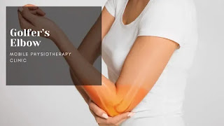
Golfer's elbow, also known as medial epicondylitis, is a condition that affects the tendons that connect the forearm muscles to the elbow. It is characterized by pain and tenderness on the inner side of the elbow, where the tendons attach to the bony bump called the medial epicondyle. 

This condition is commonly seen in people who play golf, hence the name, but can also occur in people who perform repetitive gripping activities, such as tennis players, carpenters, and painters. Symptoms of golfer's elbow include pain, tenderness, and stiffness on the inner side of the elbow, as well as weakness in the wrist and hand. 

Treatment options include rest, ice, compression, and elevation (RICE), physical therapy, and in some cases, medication or surgery.

Related Anatomy

To understand golfer's elbow, it's important to know the related anatomy. The elbow joint is formed by the humerus bone of the upper arm and the radius and ulna bones of the forearm. 

The tendons that connect the forearm muscles to the elbow joint attach to bony bumps on the humerus called epicondyles. In the case of golfer's elbow, the tendons that attach to the medial epicondyle become inflamed or damaged due to repetitive stress or overuse. These tendons are responsible for flexing the wrist and fingers and rotating the forearm, which are common movements performed in golf, tennis, and other activities that involve gripping and twisting motions. 

The muscles involved in golfer's elbow include the pronator teres, flexor carpi radialis, and palmaris longus. Other structures in the area include nerves such as the ulnar nerve and blood vessels such as the brachial artery.

Causes of Golfers elbow

Golfer's elbow is caused by overuse or repetitive stress on the tendons that attach to the medial epicondyle of the elbow. Some common causes of golfer's elbow include:

  • Repetitive gripping: Repeatedly gripping and twisting actions, such as those used in golf, tennis, or weightlifting, can strain the tendons in the forearm.
  • Improper technique: Using improper technique while performing sports or activities that require repetitive elbow movements can increase the risk of golfer's elbow.
  • Age and gender: This condition is more common in people aged 40 and above and is also more common in men.
  • Occupational hazards: Certain occupations that involve repetitive gripping or twisting actions, such as carpentry, plumbing, and painting, can increase the risk of golfer's elbow.
  • Trauma: A direct blow to the elbow or a fall on an outstretched arm can cause golfer's elbow.
  • Overloading: Overloading the forearm muscles by lifting heavy objects, performing excessive wrist curls or forearm exercises can increase the risk of golfer's elbow.

Medical conditions: Certain medical conditions, such as rheumatoid arthritis, gout, and thyroid disorders, can also increase the risk of golfer's elbow.

Symptoms of Golfers elbow

The main symptom of golfer's elbow is pain and tenderness on the inner side of the elbow, where the tendons attach to the medial epicondyle. Other symptoms of golfer's elbow may include:

  • Weakness in the wrist and hand: You may have difficulty gripping objects or performing everyday activities that require wrist or hand movement.
  • Stiffness: The elbow joint may feel stiff and may be difficult to move.
  • Numbness or tingling: In some cases, you may experience numbness or tingling in the fingers, especially the ring and little fingers.
  • Pain that worsens with activity: The pain may be mild at first but can become more severe with continued use of the affected arm.
  • Pain that radiates down the arm: The pain may radiate down the arm towards the wrist and fingers.
  • Pain when bending or twisting the wrist: You may experience pain when bending or twisting the wrist, as these movements put additional strain on the affected tendons.
  • Pain when lifting or carrying objects: You may experience pain when lifting or carrying objects, as these actions also place stress on the affected tendons.

If you experience any of these symptoms, it's important to see a healthcare provider for an accurate diagnosis and treatment plan.

Risk factor

There are several risk factors that can increase the likelihood of developing golfer's elbow. These include:

  • Age: As mentioned earlier, golfer's elbow is more common in individuals over the age of 40.
  • Gender: Males are more likely than females to develop golfer's elbow.
  • Sports participation: Individuals who participate in sports that involve repetitive gripping and twisting actions, such as golf, tennis, and weightlifting, are at increased risk.
  • Occupational hazards: Certain occupations that require repetitive gripping and twisting actions, such as carpentry, plumbing, and painting, can increase the risk of golfer's elbow.
  • Poor technique: Using improper technique while performing repetitive elbow movements can increase the risk of golfer's elbow.
  • Weak forearm muscles: Weakness in the forearm muscles can make the tendons more vulnerable to injury and increase the risk of golfer's elbow.
  • Medical conditions: Certain medical conditions, such as rheumatoid arthritis, gout, and thyroid disorders, can also increase the risk of golfer's elbow.
  • Genetics: There may be a genetic predisposition to developing golfer's elbow, although more research is needed to fully understand this connection.

It's important to note that not everyone with these risk factors will develop golfer's elbow, and some individuals may develop the condition without any identifiable risk factors.

Differential Diagnosis

There are several conditions that can cause similar symptoms to golfer's elbow. A healthcare provider may perform a thorough examination and consider other possible diagnoses before confirming a diagnosis of golfer's elbow. Some of the conditions that can be considered in the differential diagnosis of golfer's elbow include:

  • Tennis elbow: Tennis elbow, also known as lateral epicondylitis, is a condition that causes pain and tenderness on the outer side of the elbow. This condition is caused by overuse or repetitive stress on the tendons that attach to the lateral epicondyle of the elbow.
  • Cubital tunnel syndrome: Cubital tunnel syndrome is a condition that occurs when the ulnar nerve, which runs through a narrow tunnel in the elbow, becomes compressed or irritated. This can cause pain, numbness, and tingling in the elbow, hand, and fingers.
  • Radial tunnel syndrome: Radial tunnel syndrome is a condition that occurs when the radial nerve, which runs through the forearm, becomes compressed or irritated. This can cause pain and weakness in the forearm, wrist, and hand.
  • Osteoarthritis: Osteoarthritis is a degenerative joint disease that can affect the elbow joint, causing pain, stiffness, and limited range of motion.
  • Bursitis: Bursitis is a condition that occurs when the bursae, small fluid-filled sacs that cushion the joints, become inflamed. This can cause pain, swelling, and limited range of motion in the affected joint.
  • Fracture: A fracture or break in the bones of the elbow can cause pain and limited range of motion in the joint.
  • Tendinitis: Tendinitis is a condition that occurs when a tendon becomes inflamed, causing pain, swelling, and limited range of motion in the affected joint.

It's important to consult with a healthcare provider for an accurate diagnosis and appropriate treatment plan.

Diagnosis

A diagnosis of golfer's elbow is usually made based on a physical exam and a review of the patient's medical history. During the physical exam, the healthcare provider will examine the affected elbow, looking for tenderness and pain on the inner side of the elbow. They may also perform certain maneuvers to assess the strength and range of motion in the affected arm.

In some cases, imaging tests such as X-rays or MRI scans may be recommended to rule out other conditions or to further evaluate the extent of the injury. Blood tests may also be ordered to rule out other medical conditions that may be causing the symptoms.

It's important to seek medical attention if you are experiencing pain, tenderness, or other symptoms in the elbow, as these symptoms can be caused by a variety of conditions and may require different treatment approaches.

Treatment of Golfers elbow

Treatment for golfer's elbow typically involves a combination of rest, medication, physical therapy, and in some cases, surgery. The goal of treatment is to relieve pain, reduce inflammation, and promote healing.

Some common treatments for golfer's elbow include:

  • Rest: Resting the affected arm and avoiding activities that cause pain or discomfort can help reduce inflammation and promote healing.
  • Medications: Over-the-counter pain relievers such as ibuprofen or acetaminophen can help reduce pain and inflammation. In some cases, your doctor may prescribe stronger medications or corticosteroid injections to reduce inflammation.
  • Physical therapy: Stretching and strengthening exercises can help improve flexibility and reduce the risk of future injury. A physical therapist may also use techniques such as massage, ultrasound, or electrical stimulation to promote healing.
  • Brace or Splint: Wearing a brace or splint on the affected arm can help provide support and reduce stress on the affected tendons.
  • Surgery: In severe cases where other treatments have not been effective, surgery may be recommended to repair or remove damaged tissue.

In addition to these treatments, your healthcare provider may also recommend lifestyle changes such as modifying your activities, using proper form and technique during activities, and wearing protective equipment such as elbow pads or wrist braces to prevent future injury.

It's important to work closely with your healthcare provider to develop a treatment plan that is tailored to your individual needs and goals.

Physiotherapy treatment

Physiotherapy is an important part of the treatment plan for golfer's elbow. A physical therapist can help develop a personalized treatment plan that may include:

  • Range of motion exercises: Stretching and range of motion exercises can help improve flexibility and reduce stiffness in the affected arm.
  • Strengthening exercises: Strengthening exercises can help improve the strength of the muscles and tendons in the affected arm, which can help reduce pain and prevent future injury.
  • Manual therapy: Manual therapy techniques such as massage, myofascial release, and joint mobilization can help reduce pain, improve range of motion, and promote healing.
  • Ultrasound or electrical stimulation: These therapies can help reduce pain and inflammation and promote healing.
  • Ergonomic assessment: A physical therapist can assess your work environment and suggest modifications to your posture or work habits to reduce the risk of future injury.
  • Activity modification: Your physical therapist can help you modify your activities to reduce stress on the affected arm and prevent further injury.

It's important to follow your physical therapist's instructions carefully and consistently to ensure the best possible outcome. Physiotherapy is often a long-term process and may require several sessions over a period of weeks or months to achieve the desired results.

How to Prevent Golfers elbow?

There are several steps you can take to prevent golfer's elbow:

  • Warm up properly before physical activity: This can help improve blood flow to the muscles and tendons and reduce the risk of injury.
  • Use proper technique: Using proper technique and form during physical activity can help reduce stress on the tendons and muscles in the elbow.
  • Strengthen your forearm muscles: Strengthening your forearm muscles can help improve your grip strength and reduce the risk of injury.
  • Take breaks and rest: Taking frequent breaks during activities and resting the affected arm can help reduce the risk of injury and promote healing.
  • Use proper equipment: Using equipment that is properly fitted and appropriate for the activity can help reduce the risk of injury.
  • Avoid repetitive activities: Avoiding activities that involve repetitive gripping or twisting motions can help reduce the risk of injury.
  • Maintain overall fitness: Maintaining overall fitness and flexibility can help reduce the risk of injury and promote healing.

By taking these steps, you can help reduce your risk of developing golfer's elbow and other types of overuse injuries. If you do experience pain or discomfort in the elbow, it's important to seek medical attention and follow appropriate treatment recommendations.

Conclusion

In conclusion, golfer's elbow is a common condition that causes pain and tenderness on the inner side of the elbow. It is typically caused by overuse or repetitive strain on the tendons and muscles in the forearm. Treatment for golfer's elbow may include rest, medication, physical therapy, and in some cases, surgery. 

Physiotherapy is an important part of the treatment plan and can help improve range of motion, reduce pain and inflammation, and promote healing. To prevent golfer's elbow, it's important to warm up properly before physical activity, use proper technique, strengthen forearm muscles, take breaks and rest, use proper equipment, avoid repetitive activities, and maintain overall fitness. 

If you experience pain or discomfort in the elbow, it's important to seek medical attention and follow appropriate treatment recommendations.

Sunday 19 March 2023

Cervical spondylosis: Cause, Symptoms, Treatment, Exercise

 What is Cervical spondylosis?

Cervical spondylosis

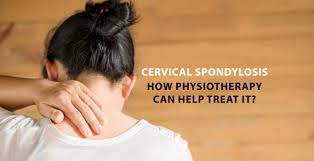
Cervical spondylosis is a medical condition that refers to the age-related wear and tear of the cervical spine (the uppermost part of the spine located in the neck). It is a degenerative condition that affects the vertebral bones, intervertebral discs, and surrounding structures such as ligaments and muscles.

The degeneration of the cervical spine can lead to the development of bone spurs (also known as osteophytes) which can impinge on nerves, resulting in pain, numbness, tingling or weakness in the arms, neck, or shoulders. Cervical spondylosis can also cause a reduction in the range of motion of the neck and can lead to headaches.

Cervical spondylosis is commonly seen in people over the age of 40, but it can occur in younger people as well, especially those who have a history of neck injuries or trauma. The condition can be diagnosed through imaging tests such as X-rays, CT scans or MRI scans, and treatment options can include physical therapy, medications, or in some cases, surgery.

Related Anatomy

The cervical spine is made up of seven vertebrae (C1 to C7) that begin at the base of the skull and extend down to the thoracic spine. The cervical spine is responsible for supporting the weight of the head, allowing for head movement and protecting the spinal cord.

Between each of the cervical vertebrae, there is an intervertebral disc, which acts as a cushion to absorb shock and prevent the vertebrae from rubbing against each other. The intervertebral discs are composed of a tough outer layer called the annulus fibrosus and a soft gel-like center called the nucleus pulposus.

The cervical spine is also surrounded by ligaments, which provide stability to the joints between the vertebrae, and muscles, which control the movement of the neck and head. The major muscles of the neck include the sternocleidomastoid, splenius capitis, levator scapulae, and trapezius muscles.

The cervical spine is also home to several important nerves, including the spinal cord, which carries signals from the brain to the rest of the body, and the cervical nerves, which branch off from the spinal cord and innervate the neck, shoulders, arms, and hands.

Causes of Cervical spondylosis

The exact cause of cervical spondylosis is not known, but it is believed to be caused by a combination of factors including age-related degeneration, genetic predisposition, and lifestyle factors. Some of the common causes of cervical spondylosis include:

Age-related wear and tear: As we age, the intervertebral discs in the cervical spine lose their flexibility and become less able to absorb shock. This can lead to the development of bone spurs or osteophytes, which can put pressure on the nerves and cause pain.

Repetitive stress: Repetitive stress on the neck, such as from poor posture or spending long hours in front of a computer, can also contribute to the development of cervical spondylosis.

Neck injuries: Trauma to the neck, such as from a car accident or sports injury, can cause damage to the cervical spine and increase the risk of developing cervical spondylosis.

Genetics: There is some evidence that genetics may play a role in the development of cervical spondylosis, as it tends to run in families.

Smoking: Smoking can accelerate the degenerative process in the cervical spine, increasing the risk of developing cervical spondylosis.

Other risk factors for cervical spondylosis include a sedentary lifestyle, obesity, and a history of heavy lifting.

Symptoms of Cervical spondylosis

The symptoms of cervical spondylosis can vary depending on the severity and location of the degenerative changes in the cervical spine. Some common symptoms of cervical spondylosis include:

Neck pain: Pain in the neck is the most common symptom of cervical spondylosis. The pain may be dull or sharp and may radiate to the shoulders, arms or hands.

Stiffness: The neck may feel stiff or rigid, making it difficult to turn the head or move the neck.

Headaches: Cervical spondylosis can cause tension headaches that start at the base of the skull and radiate to the forehead.

Tingling or numbness: Compression of nerves in the cervical spine can cause tingling or numbness in the shoulders, arms or hands.

Weakness: Compression of nerves can also cause weakness in the muscles of the shoulders, arms or hands.

Loss of balance: In rare cases, severe cervical spondylosis can cause loss of balance or difficulty walking.

The symptoms of cervical spondylosis may worsen with prolonged sitting or standing, and may improve with rest or changes in posture. It's important to seek medical attention if you experience any of these symptoms, as they can be indicative of other conditions as well.

Risk factor for cervical spondylosis

There are several risk factors that increase the likelihood of developing cervical spondylosis. These include:

Age: Cervical spondylosis is more common in people over the age of 40, as the cervical spine undergoes degenerative changes with age.

Genetics: There may be a genetic component to cervical spondylosis, as it tends to run in families.

Occupation and lifestyle: Certain occupations that involve repetitive stress on the neck, such as computer work, can increase the risk of developing cervical spondylosis. A sedentary lifestyle, lack of exercise, and obesity can also increase the risk.

Trauma: Previous neck injuries, such as those sustained in car accidents or sports, can increase the risk of cervical spondylosis.

Smoking: Smoking has been shown to accelerate the degenerative changes in the cervical spine, increasing the risk of cervical spondylosis.

Other medical conditions: Certain medical conditions, such as rheumatoid arthritis, can increase the risk of developing cervical spondylosis.

It's important to note that having one or more of these risk factors does not necessarily mean that a person will develop cervical spondylosis, but it does increase the likelihood.

Difference Diagnosis

The symptoms of cervical spondylosis can be similar to those of other conditions, so it's important to get a proper diagnosis. Some conditions that can be confused with cervical spondylosis include:

Herniated disc: A herniated disc occurs when the soft gel-like center of an intervertebral disc protrudes through the tough outer layer, putting pressure on the nerves. This can cause similar symptoms to cervical spondylosis.

Spinal stenosis: Spinal stenosis is a narrowing of the spinal canal that can put pressure on the spinal cord and nerves, causing symptoms similar to cervical spondylosis.

Pinched nerve: A pinched nerve can occur anywhere along the path of a nerve, causing pain, numbness, or weakness in the affected area.

Tension headache: Tension headaches can cause pain and stiffness in the neck, which can be mistaken for symptoms of cervical spondylosis.

Fibromyalgia: Fibromyalgia is a chronic pain condition that can cause widespread pain, fatigue, and sleep disturbances, which can be mistaken for symptoms of cervical spondylosis.

To make a differential diagnosis, a doctor may perform a physical exam, take a medical history, and order diagnostic tests, such as X-rays, MRI scans, or nerve conduction studies, to determine the underlying cause of the symptoms.

Diagnosis

To diagnose cervical spondylosis, a doctor may perform the following:

Medical history: The doctor will ask about the patient's symptoms, medical history, and any previous injuries or surgeries.

Physical exam: The doctor will perform a physical exam to assess the range of motion in the neck, strength and sensation in the arms and hands, and any signs of nerve damage.

Imaging tests: Imaging tests such as X-rays, CT scans, or MRI scans can help visualize the extent of the degenerative changes in the cervical spine.

Electromyography (EMG): EMG is a test that measures the electrical activity of muscles and nerves. This test can help determine if there is any nerve damage.

Nerve conduction studies: Nerve conduction studies are tests that measure the speed at which nerves conduct electrical signals. These tests can help determine if there is any nerve damage.

Based on the results of these tests, the doctor can make a diagnosis of cervical spondylosis and determine the severity of the condition. The doctor may also recommend a treatment plan based on the individual patient's needs.

Treatment of Cervical spondylosis

The treatment of cervical spondylosis typically depends on the severity of the condition and the individual patient's symptoms. Some common treatment options include:

Medications: Non-steroidal anti-inflammatory drugs (NSAIDs) can help relieve pain and inflammation. Muscle relaxants can also help relieve muscle spasms. In more severe cases, corticosteroids or opioids may be prescribed.

Physical therapy Treatment: Physical therapy can help improve strength and flexibility in the neck and shoulders. This can help reduce pain and improve mobility.

Neck traction: Neck traction involves the use of weights or a pulley system to gently stretch the neck. This can help relieve pressure on the nerves and improve range of motion.

Cervical collar: A cervical collar may be used to immobilize the neck and relieve pain. This is typically used for short periods of time and under the supervision of a doctor or physical therapist.

Surgery: In severe cases, surgery may be necessary to relieve pressure on the nerves. This may involve removing bone spurs, herniated discs, or other structures that are compressing the nerves.

Lifestyle changes: Making lifestyle changes such as maintaining good posture, avoiding prolonged sitting or standing, and engaging in regular exercise can help improve the symptoms of cervical spondylosis.

It's important to work with a healthcare provider to determine the most appropriate treatment plan for cervical spondylosis, as the severity of the condition can vary from person to person.

Physiotherapy treatment

Physiotherapy can be an effective treatment option for cervical spondylosis. A physiotherapist can design a treatment plan tailored to the individual patient's needs and symptoms. Some common physiotherapy treatments for cervical spondylosis include:

Manual therapy: Manual therapy techniques such as mobilization and manipulation can help improve joint mobility and reduce pain.

Exercise therapy: Exercise therapy can help improve strength, flexibility, and range of motion in the neck and shoulders. This can help reduce pain and improve mobility.

Traction: Cervical traction can help reduce pressure on the nerves and improve range of motion in the neck.

Posture correction: Poor posture can contribute to the development and progression of cervical spondylosis. A physiotherapist can help identify and correct poor posture habits.

Electrical stimulation: Electrical stimulation can be used to reduce pain and improve muscle function.

Heat and cold therapy: Heat therapy can help reduce muscle tension and improve blood flow. Cold therapy can help reduce inflammation and pain.

A physiotherapist may also provide education on self-management strategies, such as ergonomic modifications, stress management techniques, and home exercise programs. Physiotherapy can be an effective and non-invasive treatment option for cervical spondylosis, and can help improve quality of life for individuals with this condition.

Exercises for cervical spondylosis

Before beginning any exercise program for cervical spondylosis, it's important to consult with a healthcare provider or physiotherapist to determine the most appropriate exercises for your individual needs and symptoms. Some common exercises that may be recommended for cervical spondylosis include:

Neck stretches: Slowly turning the head from side to side, tilting the head forward and backward, and rotating the head in a circular motion can help improve range of motion and reduce stiffness in the neck.

Shoulder rolls: Rolling the shoulders forward and backward can help improve mobility and reduce tension in the neck and shoulders.

Chin tucks: Tucking the chin in toward the chest can help improve posture and reduce strain on the neck.

Scapular squeezes: Squeezing the shoulder blades together can help strengthen the muscles of the upper back and improve posture.

Wall angels: Standing with the back against a wall and raising the arms overhead, then lowering them back down, can help improve shoulder mobility and posture.

Resistance band exercises: Using a resistance band to perform exercises such as shoulder rows and shoulder extensions can help strengthen the muscles of the upper back and shoulders.

Isometric neck exercises
Isometric neck exercises

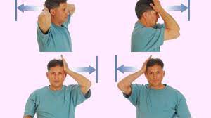
Isometric neck exercises can be a helpful addition to a comprehensive exercise program for cervical spondylosis. Isometric exercises involve contracting the muscles without actually moving the joint, which can help improve muscle strength and reduce pain. Some examples of isometric neck exercises for cervical spondylosis include:

Chin tucks: While sitting or standing, tuck your chin in toward your chest and hold the contraction for 5-10 seconds before releasing. Repeat 10-15 times.

Neck extension: While sitting or standing, place your hands behind your head and gently press your head back into your hands. Hold the contraction for 5-10 seconds before releasing. Repeat 10-15 times.

Neck flexion: While sitting or standing, place your hands on your forehead and gently press your head forward into your hands. Hold the contraction for 5-10 seconds before releasing. Repeat 10-15 times.

Lateral neck flexion: While sitting or standing, place your right hand on the side of your head and gently press your head to the right, while resisting with your neck muscles. Hold the contraction for 5-10 seconds before releasing. Repeat on the left side. Repeat 10-15 times on each side.

It's important to start slowly and gradually increase the intensity and duration of exercises over time. As with any exercise program, it's important to consult with a healthcare provider or physiotherapist to determine the most appropriate exercises for your individual needs and symptoms.

How to Prevent Cervical spondylosis?

There is no guaranteed way to prevent cervical spondylosis, as it is often a result of the natural aging process. However, there are several steps you can take to reduce your risk of developing cervical spondylosis or to prevent it from getting worse if you have already been diagnosed. These include:

Practice good posture: Maintaining good posture can help reduce strain on the neck and prevent the development of cervical spondylosis. Keep your head balanced directly over your spine, and avoid hunching forward or slouching.

Exercise regularly: Regular exercise can help keep your neck and back muscles strong and flexible, which can reduce the risk of developing cervical spondylosis.

Use proper ergonomics: Make sure your workstation is set up properly, with your computer monitor at eye level and your chair at the right height to avoid straining your neck.

Take breaks: If you spend a lot of time sitting or working at a computer, take frequent breaks to stand up, stretch, and move around.

Avoid repetitive motions: Try to avoid repetitive motions that strain your neck, such as repeatedly looking up or down or twisting your neck to one side.

Practice stress management: Stress can cause muscle tension and exacerbate symptoms of cervical spondylosis, so it's important to find ways to manage stress, such as through meditation, deep breathing, or exercise.

Quit smoking: Smoking can accelerate the degenerative process in the spine, so quitting smoking can help reduce the risk of developing cervical spondylosis or slow its progression if you have already been diagnosed.

Conclusion

Cervical spondylosis is a degenerative condition that affects the neck vertebrae, causing symptoms such as neck pain, stiffness, and numbness or tingling in the arms or hands. It is most commonly seen in individuals over the age of 50, but it can affect anyone. The condition can be diagnosed through imaging tests, and treatment options include physiotherapy, medication, and surgery.

In addition to these treatment options, there are also several ways to prevent cervical spondylosis or reduce its impact on daily life. These include practicing good posture, exercising regularly, using proper ergonomics, taking frequent breaks, avoiding repetitive motions, practicing stress management, and quitting smoking. Isometric neck exercises can also be a helpful addition to a comprehensive exercise program for cervical spondylosis, under the guidance of a healthcare provider or physiotherapist.

Overall, it's important to seek medical advice if you are experiencing symptoms of cervical spondylosis, and to take steps to prevent the condition or reduce its impact on your daily life.

Wednesday 18 May 2022

Mobile Physiotherapy Clinic Bapunagar

Mobile Physiotherapy Clinic Bapunagar
Mobile Physiotherapy Clinic Bapunagar branch is located in India colony road, Bapunagar, Ahmedabad. It is located near tollnaka char rasta opposite shaktidhara society.

Dr. Nitesh patel is a Physiotherapist and owner of the clinic and Mobile Physiotherapy clinic opened in January-2018 with purpose of quality treatment with result oriented best treatment at Home and in clinic.

Mobile Physiotherapy clinic have all the latest Physiotherapy equipment with qualified Physiotherapist, staff and located in prime area of Bapunagar.

Mobile Physiotherapy clinic Address:

B,1, Jagatnagar Society part-1, Opposite - Shaktidhara Society, India Colony Road, Tollnaka, Bapunagar, Ahmedabad, Gujarat - 380024

Website: https://mobilephysiotherapyclinic.in

Mobile no: 9499562535

Clinic timing:

Morning: 8:30am to 1:00pm

Evening: 4:00pm to 8:00pm

Sunday: 8:30am to 1:00pm

Nearby area: opposite Dinesh chambers, Near Asha Physiotherapy clinic, opposite Dr. Dinesh Patel Hospital

Physiotherapist in Mobile Physiotherapy clinic:

Dr. Nitesh Patel - Physiotherapist Mobile no- 09898607803

Dr. Priti Tirgar- Physiotherapist

Dr. Nidhi Ribadiya- Physiotherapist

Facility in The Mobile Physiotherapy clinic:

This clinic have all latest Physiotherapy equipment such as Short wave diathermy(SWD), LASER, Interferential current therapy(IFT), Transcutaneous electrical nerve stimulation (TENS), Infra-red therapy machine, Paraffin wax bath therapy, Deep heat therapy machine(DHT), Lumbar cum cervical traction machine, CPM Machine,  Electrical stimulation machine with Galvanic and faradic current.

This Electrotherapy machine are used to relive pain, swelling and muscle spasm and also useful improve mobility and reduce nerve compression.

Dr. Nitesh Patel is a Senior Physiotherapist working in Bapunagar clinic from 2005 who also running Samarpan Physiotherapy clinic near Ramji mandir India colony road.

Home Physiotherapy treatment in Bapunagar:

If you are looking for Home Physiotherapy treatment facility in Bapunagar and nearly area, Physiotherapist from Mobile Physiotherapy clinic come to your home, examine the patient and begin the treatment according to the condition of the patient.

Physiotherapy Treatment fee is affordable in Mobile clinic, mostly depends upon the condition of the patient, for that you should visit the center or you can call Dr. Nitesh Patel on 9898607803

Saturday 13 July 2019

Weight Loss And Fitness Training

Fat Loss / Weight Loss Training in Bapunagar

Weight Loss and Fitness Training is Popular nowadays's, Quality  Fitness Training Help To Improve Cardio-Vascular Blood Circulation, Improve Muscle Strength , Strengthen Joint , Increase Flexibility , Improve Pulmonary Function, Normalize Blood Sugar Level, Reduce Risk Factors Of Joint And Muscle Pain , Improve Bone Density Level, Improve Work Efficiency Level And Other Too Many Indirect Benefits Are Too.

Some Key Benefits Are As Per Below :

  • Decrease your danger of a heart attack. 
  • Deal with your weight better. 
  • Have a lower blood cholesterol level. 
  • Lower the danger of  2 -Type  diabetes and a few malignancies. 
  • Have lower Blood pressure. 
  • Decrease Mental Stress/Anxiety And Increase Dopamine Release. 
  • Exercise Reduce Chances Of Cancer. 
  • Recovering Faster From hospitalization or bed rest 
  • Have more strong bones, muscles and joints and lower danger of creating osteoporosis. 
  • Lower your Danger of falls.
Weight Loss Or Fat Loss ? :


Fat Loss / No Weight Loss


 Weight Loss By Keeping Hungry Our Body is Dangerous And There Are Too Many Side Effects , Mostly People Directly Take Action Without Proper Consulting Result In Reduce Food Intake Which Adversely Effect into Reduce Stamina, Lower Blood Pressure, And Weakness into Body, Sometimes Diabetes Patient Are Become Faintness Seen And There Are Too Many Other Side Effects. So Proper Consulting By Experts Like Dietitian, Physiotherapist, Physician Are Key To Reduce Fat Not Reduce Weight.

Fat Loss No Weight Loss Please!


Balanced Diet :
Balanced Diet Plan For Fitness/ Weight Loss


Balanced Diet Is Key in Fat Loss Programme , Balanced Diet Means Taking Proper Diet Like Eat Varieties Of Food , Have All Kind Of Vitamins, Minerals, Protein, Carbohydrate And And Minimize Fat As Much As Possible. Like Some Food Have Vitamin A But Lack Of Vitamin b Or C , So Variety Of Foods Help Provide Balanced And Each Kind Of Vitamin, Protein , Minerals, Carbohydrate And Fat And Reducing Chances Of Deficiency Disease And Make Our Body Good / Well / Fit / Strengthen. So Diet Must Of Full Of Variety With Full Of Colours Like We Seen Different Kind Of Fruits And Vegetable Are Best For Variety And Improving Body Fit And Slim With Good Amount Of Water Also. Diet Plan Change According Condition Like With Diabetes With Obesity, Or Like Joint Pain And Obesity.


Before And After Fat Loss Training


Fat Loss/ Weight Loss Training in Our Clinic : 

Obesity is a Disease As Per World Health Organization And Require Proper Treatment, And Obesity Cause Too Many Other Disease Like Coronary Artery Disease, Increase Blood Pressure, Joint Pain, Back Pain, Knee Pain, Thyroid Disease , Increase Chances Of Diabetes And Other Related Disease. So Treatment Of Obesity is Required As Early As Possible And Physiotherapist Will Help You Particularly Through Exercise And Proper Balanced Diet Plan.
Lifestyle Changes Are The Key To Reduce Weight Permanently Otherwise Losing Weight Again If Not Changes Lifestyle And Obesity is Lifestyle Disease Mostly And Somehow Heredity also.

We Provide Weight(Fat) Loss And Fitness Training in Our Clinic And at Home Also. Nearly Are Of Bapunagar, India Colony Road, Tollnka, Vastral, Amaraiwadi And Nearly Area. For Appointment Please Visit Our Contacts Us Page OR Call Us For Appointment : Dr. Nitesh Patel ( Physiotherapist ) : Mo No: 09898607803


Tuesday 9 July 2019

Paraplegia And Physiotherapy Treatment :

Paraplegia And Physiotherapy Treatment : 

 Paraplegia is paralysis or weakness (paraparesis) of both lower limbs. :

    It may be due to:
  • Pyramidal (U.M.N.) lesion resulting in spastic paraplegia.
  • Lower motor neurone (L.M.N.) lesion resulting in flaccid paraplegia.

Paralysis Treatment At Home in Ahmedabad


    It is paralysis or weakness of both lower-limbs due to bilateral pyramidal tract lesion, most commonly in the spinal cord (spinal paraplegia), and less commonly in the brain stem or the cerebral parasagittal region (cerebral paraplegia). Symptoms Mainly Unable To Walk, Stand, With Or Without Sensory Loss in Bilateral Lower Limb And With Also Affected Bladder / Bowel Incontinence.

Spinal paraplegia may be:

    1.Focal: paraplegia with sensory level.
    2.Systemic.
    3.Disseminated.


Reason Of PARAPLEGIA:

1- Focal causes:

    A. Compression:
    1. Vertebral:
Fracture or fracture-dislocation of the vertebra, Disc prolapse and spondylosis, Pott’s disease, Neoplastic 

diseases: Primary or metastatic and Deformity of the vertebral column as kyphoscoliosis.


TREATMENT  OF PARAPLEGIA: 

1. General Management:

    Frequent change of the patient’s posture to guard against bedsores.
    Care of the skin by frequent washing with alcohol followed by talc powder. In case of urinary incontinence, frequent change of  bed-sheets.
    Care of the bladder: If there is retention, use parasympathomimetic drugs. If this fails, use a catheter to evacuate the bladder.

2. Physiotherapy Treatment:
Paralysis Treatment in Bapunagar


(1) Stretching/Flexibility Exercises:
  • slow, sustained lengthening of the muscle
  • Stretching is the most important exercise you can do.
  • Stretching improves flexibility – the ability to move the parts of your body through their full range of motion.
  • Stretching also can reduce muscle spasticity and cramps and may also reduce problems such as tendonitis and bursitis.
  • To be effective, stretching routines must be done regularly, usually once or twice a day.
  • Stretch as far as you can and hold the stretch for 10 seconds and then ease back.
  • Stretching also should be done before and after other exercises to prevent muscle strain and soreness and to help avoid injuries.

(2) Aerobic Exercises:

    Steady exercise using large muscle groups
    Aerobic exercise strengthens your heart and lungs and improves your body’s ability to use oxygen. It also reduces fatigue, increases energy levels and helps you sleep better, control your weight, and lift your spirits.
    It is generally recommended to gradually work up to three or four sessions per week, each lasting 15 to 60 minutes. Include a 5-minute warm-up (including stretching) before the activity and 5 to 10 minutes of a cool down (stretching and slower activity) afterwards.
    Walking, stationary bicycling, water exercises and chair exercises are excellent choices.
    Walking: Experts recommend walking according to your ability, comfort and safety. Even short, slow walks can provide benefit.Aquatic (water) exercises: Aquatic exercises and swimming provide optimal exercise conditions. Water eliminates the effects of gravity, allowing weakened limbs to attain a greater range of motion. 

Water also helps support the body so there is less stress on hips, knees, and spine.
    Exercises in the water: Can help increase muscle power and endurance and help mobilize joints and muscles. 

They also help to relax muscles and improve coordination. Warm water (between 83 and 90 degrees F) can be especially good for stiff, sore joints. Exercises can be done while standing in shoulder-height water or while sitting in shallow water. In deeper water, an inflatable tube, floatation vest or belt can be used for flotation.
    Stationary bicycling: Stationary bicycling is a great way to improve fitness without putting stress on hips, knees, and feet. It can be done in any weather, and balance is generally not an issue. Add resistance only as you are comfortable and only after warming up.
    Chair exercises: If mobility and balance are big issues, consider chair exercises. They can provide a great workout and easily incorporate strengthening and stretching exercises.

(3) Strengthening Exercises:

    Repeated muscle contractions until the muscle becomes tired.
    Strengthening exercises help increase muscle tone and improve the quality of muscles. This enhances mobility and provides energy and a positive sense of well-being.
    Strong hip and leg muscles are needed to lift the legs to walk, and strong arm muscles are needed to carry out daily functions. Strong abdominal and back muscles help maintain correct posture and can counter pain resulting from poor gait, poor posture or the use of mobility aids.

Electrotherapy Treatment :

    Thermotherapy: are used to decrease the pain and spasticity. 
They are (i) Superficial heat:IRR, wax bath, etc. 

(ii) Deep heat: SWD, MWD, etc.
    Electrical therapy: is used to increase muscle power and to decrease the pain. e.g, TENS, EST, IFT.

Physiotherapy Technique  :Useful in Improving Condition Are : 
  • Mat exercise.
  • PNF exercise.
  • Active and passive ROM exercise.
  • Strengthening exercise.
  • Stretching exercise.
  • Endurance exercise
  • Co-ordination exercise.
  • Pelvic tilting exercise.
  • Hamstring muscle stretching.
  • Spinal rotation.
  • Calf muscle stretching.
  • Neck raising exercise
  • Knee rolling exercise.
  • Lying in extension.
  • Extension exercise.
  • Back and gluteal exercise.
3 . Symptomatic Treatment:
  • Analgesics and sedatives for pain.
  • Muscle relaxants for the spasticity.
  • Vitamins and tonics
4. Orthosis:
  • Various orthosis are used to assist patient with paraplegia.
  • These are: crutch, walker, cane, brace and wheelchair.
5. Gait training:

    It is the important part of rehabilitation program balance can be achieved by proper gait training. Gait training can be done by following methods:
    Pre ambulation MAT program:
    Rolling, prone on elbow, prone on hand, quadruped, pelvic tilting, setting and standing balance.
    Parallel bar progression
    Advanced parallel bar activities.
    Assistive device: E.g, Cane, crutches, walker

6. Home program and Ergonomics:

    Patient is advised to use the lumbosacral orthosis to support the back during traveling.
    Patient is advice for hot fomentation at home.
    Patient is advised to lying in prone position for at least 15 minutes duration twice in a day.
    Patient is explained about the proper sitting, standing, lying and lying to standing , doing the household activity in a proper way.
    patient is advised to take rest and to avoid the forward bending as much as the patient can avoid.

Patellofemoral Pain Syndrome (PFPS) : Cause, Symptoms, Treatment

What is Patellofemoral pain Syndrome? Patellofemoral pain Syndrome (PFPS) Patellofemoral pain syndrome (PFPS) is a condition characterized b...